Dark field imaging
Grating-based X-ray interferometry is a promising imaging technique that simultaneously provides attenuation, phase, and visibility contrast (dark-field) images. X-ray dark-field (DF) imaging, the subset of this technique, shows unique advantages and precision in detecting fine structures, such as micro-calcifications on Mammography and lung alveoli. However, the practical feasibility of monochromatic sources in such interferometer-based setups is severely limited in clinical and laboratory settings.
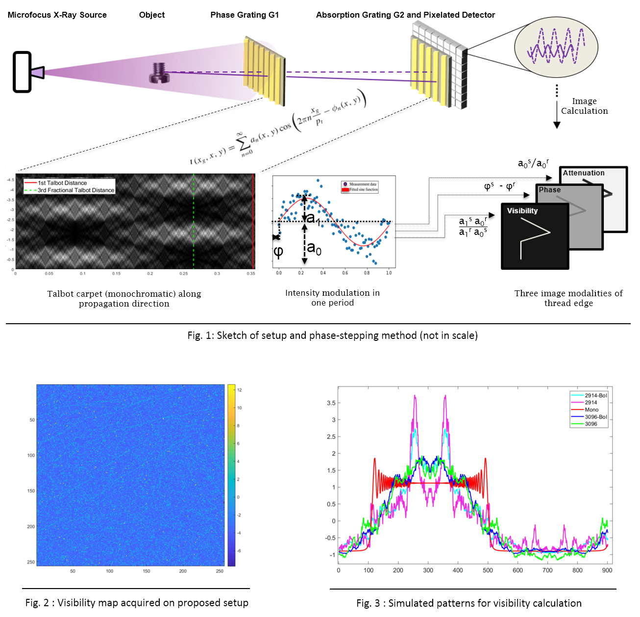
In this project, we aim to investigate the feasibility of providing a simplified compact interferometer-based X-ray DF imaging system that matches the feasibility in clinical and experimental tasks (see the upper row of Fig. 1). We further explore the proposed setup's detection limitation to provide a novel hybrid imaging tool integrating DF and XFCT for in vivo tumor profiling (IMMPRINT).

Utilizing the Fourier decomposition principle (refer to the bottom row of Fig. 1), we have observed an evenly distributed visibility map (Fig. 2) on the proposed DF imaging setup. We have determined the comparative ratio of visibility values between measurements and simulations (Fig. 3), affirming the feasibility of our compact setup for DF imaging.
With the initial measurement data, we focus on further investigating and enhancing the compact setup by developing image analysis and optimization methods. Thesis/research project topics for bachelor and master students in medical systems engineering are available. If you are interested, please contact us by e-mail and provide the following information: a short CV, transcript of academic records, areas of interest, and desired starting date.)
This project is co-related to project IMMPRINT.
