X-ray Dark Field Imaging
Recent studies in Grating-based X-ray interferometry imaging have shown promising applications in medical diagnostics. By utilizing the wavelike behavior of X-rays through a grating-based interferometer setup, various imaging modalities (attenuation contrast, phase contrast, and visibility contrast) can be obtained in a simultaneous radiation and data process. Enhanced radiographic images, such as microstructures of objects below the detector resolution, have been successfully reported in the last decade.

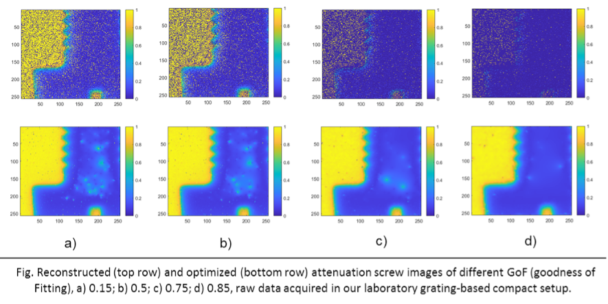
However, due to the high precision calibration requirements of optical devices in wave-based imaging systems, utilization of either the coherence properties of synchrotron sources or the high-flux capabilities of selected tabletop x-ray tubes with source gratings to achieve successful imaging has been employed in previous studies. To ease the utility in clinical and laboratory settings, we aim to build a new method of compactly combining energy-resolving x-ray detectors with polychromatic microfocus x-ray source in a Talbot-interferometer setting. With the initial measurements on a finely threaded screw, currently we challenge the data processing on image and system optimization. Hence, the following sub-projects are available for students as research or thesis topics:
1. Investigation of system parameters, including setup geometry, X-ray tube filtration, corresponding photon energy spectrum, and study/testing of phase-scan methods.
2. Image analysis and optimization, encompassing CNR calculations under various conditions (e.g., raw data re-bin and image reconstruction).
These topics can be adjusted or combined based on the interests and experience of the student. If you are interested, please contact us via email and provide a short CV, transcript of academic records, areas of interest, and desired starting date.
For more information please contact M.Sc. Xiaolei Yan


